
|
|
Radial Tears | Rim Lesions | HIZ Sign | Anular Tear Home
Concentric Anular Tears (aka: circumferential tears)
 And finally we come to the last type of anular tear. Concentric
anular tears are a separation or splitting apart of the
anulus fibrosus, between the lamellae. Figure #1 show
concentric tears (red) from a sagittal view. Concentric tears are frequently
seen in the middle and outer 1/3 of the anulus and rarely seen in the
inner anulus. It is believed that trauma is the cause of concentric
tears, especially from torsion over-load injuries, such as swinging a
golf club or throwing a discuss. Since the
outer 1/3 of the anulus is well innervated (has pain fiber and can feel)
a disruption in this region can be extremely painful!
And finally we come to the last type of anular tear. Concentric
anular tears are a separation or splitting apart of the
anulus fibrosus, between the lamellae. Figure #1 show
concentric tears (red) from a sagittal view. Concentric tears are frequently
seen in the middle and outer 1/3 of the anulus and rarely seen in the
inner anulus. It is believed that trauma is the cause of concentric
tears, especially from torsion over-load injuries, such as swinging a
golf club or throwing a discuss. Since the
outer 1/3 of the anulus is well innervated (has pain fiber and can feel)
a disruption in this region can be extremely painful!
Research has indicated that for reasons yet unknown, the lamellae lose their ability to adhere or ‘stick together’. This loss of adhesion between the lamellae is called ‘delamination’. In 1997, Vernon-Robert et al. conducted one of the very few animal studies on induced concentric anular tears. They discovered that ‘delamination’ always proceeds a full-blown concentric tear (6). It is still unclear whether this process is the result of natural aging, pathological degeneration, trauma, or a combination of each.
Interestingly, animal studies have found that induced concentric tears do NOT stimulate severe disc degeneration like rims lesions do (26). Concentric tears do however cause marked biomechanical problems within the disc and lead to thickening of the lamellae which in turn ‘stiffens’ the joint. In 2001, Fazzalari et al. demonstrated that an induced concentric tear will cause a marked loss of motion in that motion segment and cause subchondral bone thickening adjacent to the tear (26).
Other research indicates that these tears do not seem to be associated with aging or degeneration, for they are seen in equal numbers in both the young and the old (3). These tear are the most commonly seen anular tear (6) and are seen in both the anterior and posterior regions of the disc with equal frequency; except at L5/S1 where they are more common in the posterior of the disc (3).
 Concentric
tears are highly related to HIZ,
according to Ito et al (19). They noted that HIZ (high intensity zone)
is produced when a full thickness radial tear coalesces with a concentric
tear, in the very outer periphery of the posterior anulus. The radial
tear allow some nuclear material to inter into the outer anulus and set
up and inflammatory reaction. This inflammatory reaction in combination
with newly formed, vascularized granulation tissue, results in the MRI
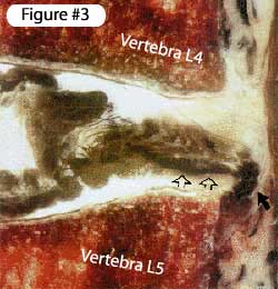
finding for HIZ. Figure #3. shows a sagittal view of
what an HIZ sign looks like histologically speaking. Note the radial tear
(white arrows)coalescing with a concentric tear (black arrow) in the very
outer periphery of the disc. There is no disruption of the PLL so this
is not an extrusion. (Ito also notes that since some rim lesion are indistinguishable
from concentric tears, rim lesion may also combine with radial tears to
form the HIZ sign.) (19) Although HIZ is not as good as a predictor at
that disc being concordantly painful on discography, it is a good screening
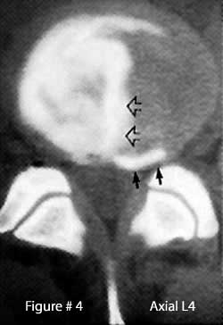
tool (19). Figure #4. is what a real HIZ looks like on
a CT discography. The black arrows point to the concentric anular tear
in the periphery of the anulus. The white arrows points to the central
radial tear.
Concentric
tears are highly related to HIZ,
according to Ito et al (19). They noted that HIZ (high intensity zone)
is produced when a full thickness radial tear coalesces with a concentric
tear, in the very outer periphery of the posterior anulus. The radial
tear allow some nuclear material to inter into the outer anulus and set
up and inflammatory reaction. This inflammatory reaction in combination
with newly formed, vascularized granulation tissue, results in the MRI
finding for HIZ. Figure #3. shows a sagittal view of
what an HIZ sign looks like histologically speaking. Note the radial tear
(white arrows)coalescing with a concentric tear (black arrow) in the very
outer periphery of the disc. There is no disruption of the PLL so this
is not an extrusion. (Ito also notes that since some rim lesion are indistinguishable
from concentric tears, rim lesion may also combine with radial tears to
form the HIZ sign.) (19) Although HIZ is not as good as a predictor at
that disc being concordantly painful on discography, it is a good screening
tool (19). Figure #4. is what a real HIZ looks like on
a CT discography. The black arrows point to the concentric anular tear
in the periphery of the anulus. The white arrows points to the central
radial tear.
Radial Tears | Rim Lesions | HIZ Sign | Anular Tear Home
References:
3) Osti OL, Vernon-Roberts B, et al. “Annular Tears & Disc Degeneration” J Bone Joint Surg [Br] 1992; 74-B:678-82
6) Vernon-Robert B, et al. “Pathogenesis of Tears of the Anulus”, - Spine 1997; 22(22):2641-46
19) Ito M, et al. “Predictive signs of discogenic lumbar pain on MRI with Discography correlation.” – Spine 1998; 23:1252-8
26) Fazzalari NL, et al. “Mechanical & Pathologic Consequences of Induced Concentric Anular Tears” – Spine 2001; 26(23):2575-2581
© Copyright 2002 – 2005 by Dr. Douglas M. Gillard DC - All rights reserved