MRI Quiz Answers:
MRI Quiz Answers:
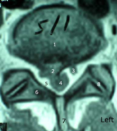
#1) L5 intervertebral disc.
#2) 9 millimeter disc herniation.
#3) Left S1 nerve root.
#4) Thecal Sac.
#5) Epidural space.
#6) Right lamina.
#7) Spinous process.
#8) Left facet joint.
Question #2) Name me the location of this 'disc lesion' using the correct zone. This is 9 mm right paracentral disc extrusion (uncontained herniation) within the right lateral recess.
Question #3) Which nerve root is being displaced by this disc lesion? The right S1 nerve root. Its also compressing the right anterolateral aspect of the thecal sac.
Question #4) bonus: What type of MRI image are we looking at??
hint: T1, T2, or Proton density?? This is a T1 Weighted scan.
Question #5) What two structures are being contacted by this disc lesion? The right S1 nerve root and the thecal sac (dura mater).
Official MRI Report on this patients:
1. At L5-S1, there is moderate disc space narrowing and moderate to marked disc desiccation with mild annular disc bulging/diffuse end plate spurring and a superimposed large central/right paracentral disc extrusion measuring up to 9 mm in anteroposterior thickness apparently representing significant enlargement of this herniation in comparison to the previous lumbar spine MRI dated 10/15/96, in which the herniation at this level measured approximately 4 mm. There is moderate to marked encroachment upon the central right S1 nerve root distal to its origin from the thecal sac, possibly with mild posterolateral displacement of the nerve root. In addition, there is a slight impression upon the right anterior aspect of the thecal sac with overall no significant central stenosis. There is no significant left lateral recess encroachment and only mild bilateral foraminal narrowing.
HISTORY:
 I saw Javier in late 1995 for a work related accident. Because of positive neurological finding an MRI and EMG were ordered. A 6 mm disc herniation at L4/5, and a 4 mm disc herniation at L5/S1 were discovered, along with a positive EMG for L5 radiculopathy. He was successfully treated with Chiropractic Cox Flexion / Distraction, PT, epidural injections, NSAIDS, and Exercise. He made a full recovery after one year.
In mid 2003, Javier was again injured doing heavy lifting while at work (something that he was told never to do again I might add!). This time, he has not been so lucky with our conservative care and epidural steroids injections and is scheduled for surgery in March. (It's unfortunate that California's Work Comp system moves at such a 'snails pace' for we have been waiting months to get this surgery authorized!) So much for the 3 month rule! (here)
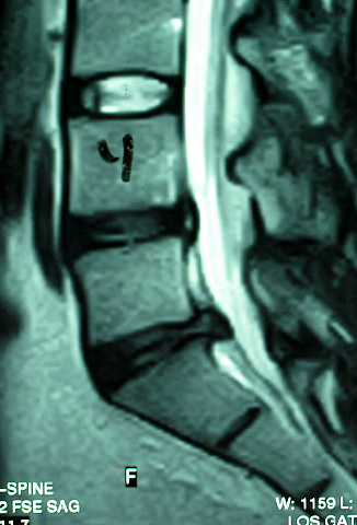
I've included his Lateral T2-weighted view for completeness. Note the moderate to severe DDD at L4 and L5. Also note the 4 mm contained herniation at L4/5, and the extrusion (non-contained herniation) at L5/S1. He has been miserable for months awaiting industrial authorization for surgery. Our care only gives him temporary pain relief. Please note that only gentle Cox flexion distraction is being used, with PT. It would be extremely dangerous for this patient to receive regular grade 5 osseous manipulation!
© Copyright 2002 – 2005 by Dr. Douglas M. Gillard DC - All rights reserved